Alzheimer’s disease remains one of the most formidable medical challenges of our time, serving as the leading cause of dementia worldwide. A critical bottleneck in patient care is that by the time clinical symptoms—such as severe memory loss and cognitive decline—manifest, billions of neurons have already suffered irreversible damage.
According to foundational scientific consensus regarding Alzheimer’s pathogenesis (as documented in PMC8371153), the disease is a decades-long, silent process orchestrated by two definitive biological footprints: Amyloid-beta (Aβ) and Tau protein. Understanding how this duo ravages the brain is vital to embracing the new era of early diagnostic screening.
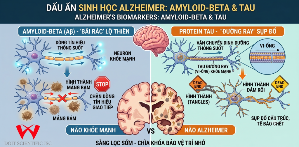
1. Amyloid-beta (Aβ): Extracellular Roadblocks in the Neuronal Network
Amyloid-beta peptides are fragments derived from the cleavage of a larger membrane protein known as APP (Amyloid Precursor Protein). In a healthy brain, these fragments are efficiently cleared and degraded.
However, the pathological cascade of Alzheimer’s disrupts this clearance mechanism. Extracellular Aβ peptides begin to aggregate, forming insoluble structures known as amyloid plaques within the synaptic spaces between neurons. The accumulation of these plaques acts like systemic roadblocks:
- They physically disrupt the synaptic transmission of electrochemical signals between brain cells.
- They trigger a chronic neuroinflammatory response from microglia, inadvertently accelerating local tissue damage.
2. Tau Protein: Intracellular Collapse of the Transport Machinery
While Amyloid-beta disrupts cellular networks from the outside, Tau protein acts as an internal agent of destruction within the neuron itself.
Under physiological conditions, neurons rely on an internal structural skeleton called microtubules—akin to a molecular railroad system—to transport essential nutrients and molecules. Tau proteins serve as the stabilization ties that keep these tracks straight and secure.
In the Alzheimer’s brain, hyperphosphorylation causes Tau molecules to detach from the microtubules and bind to one another, forming twisted structures known as neurofibrillary tangles (NFTs). The consequences are catastrophic:
- The internal transport infrastructure completely collapses.
- Deprived of essential nutrients and structural integrity, the neuron undergoes metabolic failure and eventually dies (apoptosis).
3. The Diagnostic Revolution: Identifying Biomarkers in CSF and Blood
Historically, a definitive diagnosis of Alzheimer’s was reserved for post-mortem autopsy or through highly expensive, specialized Positron Emission Tomography (PET) imaging. Today, translating clinical research into biomarker applications has introduced two transformative diagnostic paradigms:
- Cerebrospinal Fluid (CSF) Analysis: Because CSF bathes the central nervous system, a distinct profile—characterized by decreased Aβ42 levels (trapped in cerebral plaques) and elevated total Tau or phosphorylated Tau (p-Tau) levels—serves as an exceptionally accurate indicator of active pathology.
- Blood-Based Biomarkers (The Future of Screening): Thanks to ultra-sensitive immunoassay platforms (such as Simoa and advanced Mass Spectrometry), clinicians can now detect minute quantities of brain-derived p-Tau and Aβ crossing the blood-brain barrier. Blood testing marks a paradigm shift, offering a scalable, non-invasive, cost-effective tool for routine population screening.
Conclusion
The interplay between Amyloid-beta plaques and Tau tangles represents the core engine of Alzheimer’s progression. Capturing these biological footprints during the preclinical stage—years before cognitive deficits emerge—provides an invaluable window for targeted therapeutic interventions, lifestyle modifications, and cognitive preservation.
At DOIT Scientific, we stand at the forefront of biotechnology, delivering advanced screening frameworks to ensure that Alzheimer’s disease is caught early, protecting the memories of generations to come.